Diagnostic Imaging Technologies
DMC imaging technology offers some of the latest in imaging technology, allowing physicians to
“see” into the body to diagnose diseases and injuries before developing treatment plans. With advanced imaging technologies on site and expert subspecialty radiologists available 24 hours a day, we can provide expert interpretation of
images.
Diagnostic imaging technologies and interventional procedures offered by DMC are below. Looking for a specific test or imaging procedure? Call the DMC Health Access Center at 888-DMC-2500 for help finding the test you need.
Bone Density Scanning
Bone density generally decreases as we age, and for women it often drops shortly after menopause when declining estrogen levels no longer protect bone density. Older men are also at risk for lower bone density and osteoporosis,
as are people who have taken medications that affect bone density for prolonged periods (such as some asthma and steroid medications) and people with a history of osteoporosis or fractures in their family.
A bone density test determines your
T-Score, which compares your bones to healthy bones. Knowing your T-Score and risk factors helps you and your physician decide how aggressively you should address osteoporosis prevention and treatment.
Computed Tomography (CT) Scan
CT scanners are capable of capturing numerous wafer-thin images of the body within seconds.
Using computer technology, the images of your body may be viewed and manipulated in multiple projections to more clearly visualize the internal organs and structures. The equipment also allows for a wider range of clinical exams, faster exam times and
increased patient comfort.
CT has several advantages over a standard x-ray. It can distinguish between different types of soft tissue (showing a difference between the liver and the kidneys, for example). In addition, CT Scans can be combined to produce a three-dimensional image,
which can provide additional insight to the affected areas.
For your safety, please inform the CT staff if you are or have:
- An allergy or previous reaction to iodine or x-ray contrast
- History of kidney disease
- Sickle cell disease
- Pheochromocytoma
- Multiple myeloma
- Pregnant or breastfeeding
- Diabetic and taking Metformin, Glucophage, or similar medications
Mammography and Breast Imaging
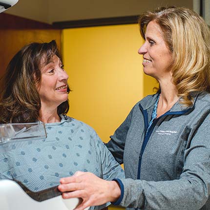
A Mammogram is an x-ray that images inside the breast. Our breast imaging services include screening and diagnostic mammograms, computer aided detection, cyst aspiration, ultrasound guided needle biopsy, stereotactic guided biopsy, needle localization
and ductogram.
Magnetic Resonance Imaging (MRI)
DMC radiologists use magnetic resonance imaging (MRI) to diagnose and treat many medical conditions. MRI uses a powerful magnetic field to produce detailed images of organs, soft tissues, bone and virtually all other internal body structures. Detailed
MRI scans allow physicians to better evaluate various parts of the body and determine the presence of certain diseases that may not be seen with other imaging methods such as X-rays, ultrasound or computed tomography (CT).
MRI does not use X-ray radiation, instead using the millions of atoms in the body are magnetic to create pictures. When placed in the MRI, these atoms line up, much like a compass points to the North Pole. Radio waves, tip these tiny magnets away
from the magnetic field. When the radio waves are turned off, the atoms move back. A powerful antenna picks up these signals and sends them to a computer that produces a black-and-white image.
MRI can provide valuable information on many conditions. In some cases, a specialized MRI can be a helpful tool in diagnosing disease. This is especially important when other methods of imaging are unable to provide the information your physician
needs to make a diagnosis or develop a treatment plan. Specialized MRI scans offered at DMC include a Breast MRI, Cardiac MRI, Diffusion Tensor Imaging (DTI), MR Angiography (MRA), MR Arthrography and MR Perfusion Exams.
Most MRI machines are a tube, which can make you feel uncomfortable if you are claustrophobic or don’t like tight spaces. In order to better serve our community, DMC has invested in an Open-Bore MRI, available at our Midtown Detroit campus.
This revolutionary scanner features an extra-large opening and is only four feet long, allowing patients weighing up to 500 pounds or those who may experience claustrophobia can get the results they need with ease.
For your safety, please notify the MRI staff if you have:
- A cardiac pacemaker
- An artificial heart valve
- A cerebral aneurysm clips
- A history of open-heart surgery
- A history of or presently have metal fragments in your eyes
- A history of chronic renal failure or are on renal dialysis
- A metal plate, pin or other metallic implant
- An intrauterine device, such as a Copper-7 IUD
- A neurostimulator
- An ear prosthesis/cochlear implant
- Permanent (tattoo) eyeliner
- A previous gunshot wound, shrapnel
- Pregnant or breastfeeding
- Claustrophobic
- Unable to lie flat or still for any length of time
Nuclear Medicine (SPECT/CT)
DMC Nuclear Medicine specialists use a variety of advanced procedures to help diagnose and treat diseases. Unlike traditional imaging procedures that focus on the structure of organs, tissue and bones, nuclear medicine evaluates the function of organs, tissue and bones.
During these procedures, small amounts of radioactive material are injected to help special cameras take images of the radioactive material as it decays and moves through targeted body processes and organ functions.
SPECT/CT is a combination of a SPECT (Single Photon Emission Computer Tomography) scan with a CT (Computed Tomography) scan. A SPECT/CT scan uses a radioactive material and a special camera to create 3D images. While most imaging tests show what internal organs look like, SPECT/CT can show how well organs are working such as blood flow, activity in the brain, etc.
Positron Emission Tomography/CT (PET/CT) Scan
Positron Emission Tomography/CT (PET/CT) scans can effectively identify many of the most common cancers, heart diseases and nerve diseases before they can be seen on other tests. PET/CT scans are particularly useful for diagnosing and treating
a range of diseases and conditions, including many types of cancer, tumors of the muscles and bones, some cardiovascular or neurological conditions and more.
PET/CT may enable doctors to:
- See tiny tumors and allow for an early diagnosis
- Determine how far cancer, heart and neurological diseases have progressed
- Identify diseased tissue, cancerous tissue and scar tissue
- Avoid performing more-invasive exams or surgeries
- Assess or "stage" cancer prior to surgeries or treatment
- Determine the effectiveness of a therapy
These scans are able to show physicians the difference between healthy tissue and diseased tissue, cancerous tissue and scar tissue. Since these changes in the way cells function often occur before a physical change can be found, PET/CT may help your
doctor make an earlier diagnosis. This can result in faster, more effective treatment and may help to avoid other exams or surgeries.
Prior to your scan, a small amount of FDG, a radioactive sugar tracer, will be injected into your hand or forearm. FDG has no known side effects and leaves your body when you urinate. It takes 45 to 60 minutes for the FDG tracer to concentrate in
the tissue so a proper scan can be made. Once the FDG has moved into your cells, you will be positioned on a comfortable table that moves slowly through the PET/CT scanner. The system has spacious, well-lit openings on both ends, and the machine
will make noise during the scan. To help you relax, you may listen to music or use ear plugs.
Radiology
DMC Radiologists use a variety of radiology procedures to diagnose and treat patients. Some specialty radiology techniques include fluoroscopy and arthrography.
Fluoroscopy
Fluoroscopy is an imaging technique used for obtaining real-time, moving X-ray images of a patient. Think of it as an X-ray TV camera that allows the radiologist to watch live images on a TV monitor. It is often used to visualize the digestive tract
and to observe surgical instruments during minimally invasive procedures.
Arthrography
Arthrography is a type of X-ray procedure used to examine a joint when standard X-rays are not adequate. A series of X-rays is taken with the joint in various positions after a contrast agent is injected into the joint. It is commonly used to examine
the knee, shoulder and wrist.
Ultrasonography (Ultrasound)
DMC experts use advanced ultrasonography equipment to capture real-time images of internal organs, muscles and tendons. Our highly experienced physicians and ultrasound technologists use ultrasound as a diagnostic imaging tool and as guidance for
needle biopsies of tumors and therapeutic drainages of fluid collections.
Ultrasound technology is often used for abdominal, gynecologic and small body part assessment. Abdominal ultrasounds can look for liver disease, kidney disease, aortic aneurysms, gall bladder conditions, organ transplant health, pancreas disease and
more. For small body parts, ultrasound can help identify thyroid tumors and conditions, muscle & tendon issues, prostate tumors and more.
Perhaps the most common association with ultrasounds is their use to monitor a fetus while in the mother’s womb. Gynecologic ultrasounds can do more than that, also identifying uterine fibroids, endometriosis, ovarian tumors, ectopic pregnancy
and more.
X-ray
X-rays were the first type of medical imaging technology to be developed, and are useful in identifying bone and even some soft tissue diseases. One common example is the chest X-ray, which can identify lung diseases such as pneumonia, lung cancer
or pulmonary edema. However, traditional X-rays are not useful in the imaging of most soft tissues such as the brain or muscle. Imaging alternatives for soft tissues are computed tomography (CT scanning), magnetic resonance imaging (MRI) or ultrasound.


